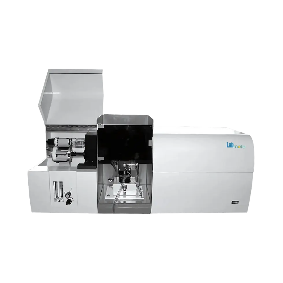
Labmate Microscope Camera LMMIC-A108
Labmate Microscope Camera LMMIC-A108 delivers 4.0 MP high-definition imaging with smooth 30 fps video capture. It supports manual and auto exposure modes, progressive scanning, and electronic shutter control for clear and precise imaging. HDMI and dual USB 2.0 interfaces enable easy connectivity and storage, while adjustable cross lines and built-in measurement tools enhance analytical accuracy. The camera also includes freeze function for split-screen comparisons.
Product Info
Key Features
-
4.0 MP SONY color CMOS sensor
-
30 fps real-time video
-
Progressive scanning mode
-
Manual and auto exposure
-
Electronic shutter control
-
Auto/manual white balance adjustment
-
Adjustable cross lines (1–12 groups, configurable size and color)
-
Freeze function with two or four split screens
-
Built-in measurement tools: distance, rectangle, circle, angle
-
HDMI digital output
-
USB storage support for images and videos
Technical Highlights
Specification Details Sensor Type 4.0 MP, 1/3” SONY color CMOS Effective Pixels 2560H × 1440V Data Bits 12 bit Pixel Size 2.0 µm × 2.0 µm Frame Rate 30 fps Definition >1200 TV lines Scanning Mode Progressive Shutter Mode Electronic shutter Dynamic Range >90 dB Signal-to-Noise Ratio 55 dB Sensitivity 2.0 V/lux-sec (550 nm) Exposure Mode Manual / Auto White Balance Auto / Manual Lens Mount C / CS Output Interface HDMI, 2 × USB 2.0 HDMI Output Resolution 1920 × 1080 / 2560 × 1440 Light Source DC 12V / 1A Dimensions 53 × 53 × 45 mm Menu Function USB interface for mouse/U disk, adjustable cross lines, freeze function, measurement tools Accessories
Standard Accessories
-
HDMI cable (1.5 m) – 1 pc
-
USB storage – 1 pc
-
Power adapter (DC 12V / 1A) – 1 pc
-
Lens cleaning cloth – 1 pc
-
Calibration slide – 1 pc
-
Microscope mount adapters – 2 pcs
-
Protective carrying case – 1 pc
-
User manual – 1 pc
Applications
Labmate Microscope Camera LMMIC-A108 is ideal for clinical diagnostics, forensic analysis, industrial quality control, biological research, pathology, cytology, material inspection, and live-cell imaging, providing high-definition and accurate imaging for detailed analysis.-